

What Does Skin Cancer Look Like? Skin cancer is one of the most prevalent types of cancer affecting millions of people worldwide each year, and the scary truth is that many individuals don’t recognize the early warning signs of skin cancer until it’s too late. If you’ve ever noticed an unusual skin lesion on your body or spotted mole changes that seemed out of the ordinary, you’re not alone in your concern. The good news is that skin cancer, when caught early through regular dermatologist exam and self-monitoring, has a significantly higher survival rate. This comprehensive guide will walk you through exactly what skin cancer looks like, how to identify different types of cancer including melanoma, basal cell carcinoma, squamous cell carcinoma, and other dangerous growths. Whether you’re worried about a new pigmented lesion, a non-healing sore, or a mysterious scaly patch on your skin, understanding the visual characteristics and recognizing warning signs using the ABCDE rule can be your first line of defense in protecting your health. Most skin cancer cases develop due to sun exposure and the damaging effects of UV radiation, but knowing what signs to look for gives you the power to take action before serious complications arise.
The Three Main Types of Skin Cancer and Their Appearance
Melanoma: The Most Aggressive Form of Skin Cancer
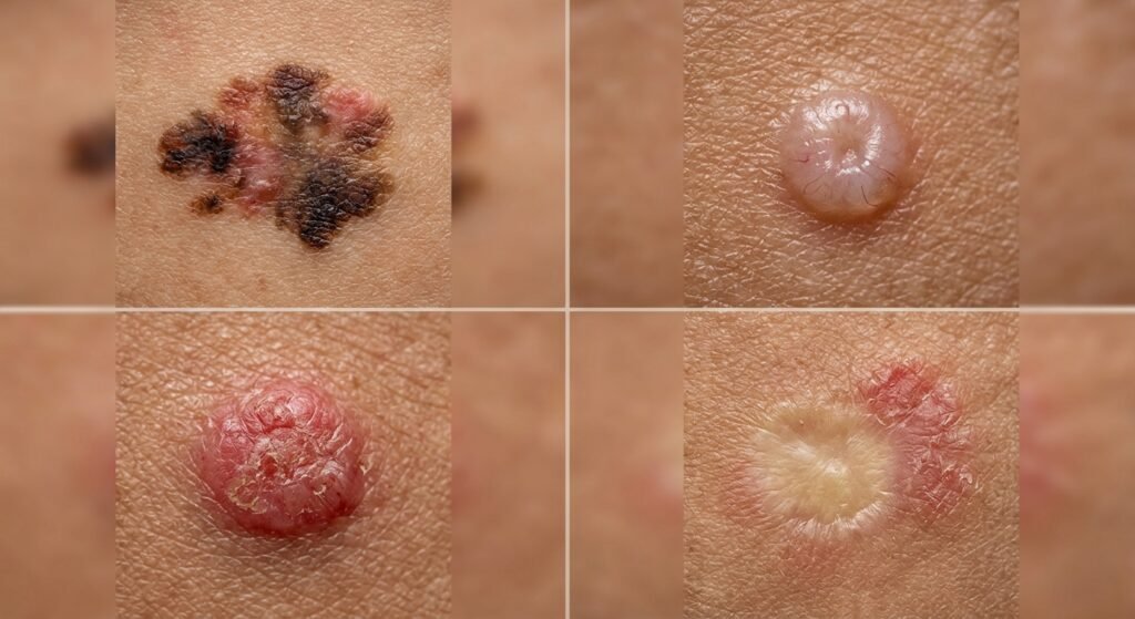
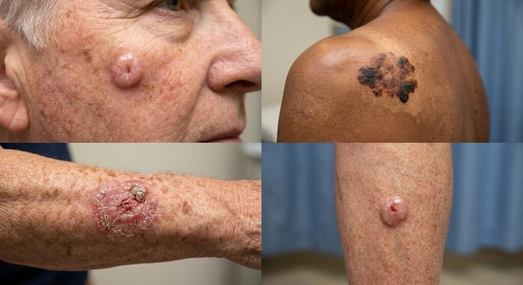
When dermatologists talk about the most dangerous type of skin cancer, they’re referring to melanoma. This aggressive form of cancer develops in the melanocytes, the cells responsible for producing melanin, the pigment that gives your skin its color. What makes melanoma particularly concerning is that it can spread rapidly to other parts of your body if not treated early. A melanoma often appears as an irregularly shaped mole that looks distinctly different from your other moles. You might notice that it has multiple colors—perhaps browns, blacks, reds, or even blue tones all mixed together in one area. The borders are typically jagged and irregular rather than smooth and well-defined like a normal mole should be.
One of the most telling signs of melanoma is when a mole starts changing. If you’ve had the same mole for years and suddenly it begins to grow larger, change color, or develop an uneven appearance, this warrants immediate attention from a dermatologist. Melanoma lesions tend to be larger than six millimeters in diameter, which is roughly the size of a pencil eraser. The pigmented lesion may also develop asymmetry, meaning one half doesn’t match the other half. Some people describe a concerning melanoma as looking like a dark, irregular spot that seems to be spreading across the skin in an uncontrolled manner.
Basal Cell Carcinoma: The Most Common but Often Overlooked Type
Basal cell carcinoma accounts for about eighty percent of all skin cancer cases, making it the most frequently diagnosed type. Despite being common, many people overlook or misidentify basal cell carcinoma because it doesn’t always look like what they expect cancer to look like. This type of skin cancer typically appears as a shiny bump on the skin, often with a translucent or pearl-like quality that can seem almost innocent to the untrained eye. The bump might be pink, red, or even skin-colored, and it frequently has a small indentation or dimple in the center.
As basal cell carcinoma develops, it can take on different appearances. Some lesions look like a pink growth with a slightly raised border, while others develop a scar-like appearance that looks remarkably like a scar from an old injury. You might notice tiny blood vessels visible on the surface of the lesion, or there might be areas of crusting or bleeding. The skin lesion tends to grow slowly, which is why many people don’t realize they have a problem until it’s been there for quite some time. Basal cell carcinoma typically appears on areas of the skin that receive the most sun exposure, particularly the face, ears, scalp, and neck.
Squamous Cell Carcinoma: The Scaly and Persistent Threat
Squamous cell carcinoma is the second most common type of skin cancer, and it has some distinctive visual characteristics that can help you identify it. This type of lesion often presents as a scaly patch that feels rough or crusty to the touch. The affected area might appear red, inflamed, or irritated, and the scaling can resemble eczema or dermatitis at first glance, which is why it’s sometimes initially misdiagnosed. Some squamous cell carcinomas develop as wart-like growths that are raised above the surrounding skin surface.
One of the hallmark features of squamous cell carcinoma is the appearance of a sore that doesn’t heal. If you notice any area on your skin that seems to be an open wound, crust, or ulcer that persists for more than three weeks without showing signs of improvement, this is a red flag that demands professional evaluation. These non-healing sores often bleed easily and may have a tender or painful quality. The mole changes associated with squamous cell carcinoma tend to happen on sun-exposed areas, with the face, lips, ears, hands, and forearms being the most common locations where this cancer develops.
Other Important Types of Skin Cancer You Should Know About
Actinic Keratosis: The Precancerous Warning Sign
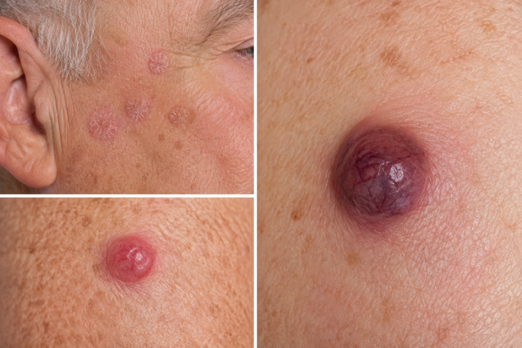
Actinic keratosis, sometimes called solar keratosis, represents a precancerous condition that many dermatologists treat seriously because of its potential to progress into squamous cell carcinoma. These lesions appear as small, scaly patches on the skin, typically rough to the touch and irregular in texture. You might describe the feeling as similar to sandpaper or a callus. Actinic keratosis patches are usually red, brown, tan, or flesh-colored, and they tend to appear in clusters on areas that have experienced significant sun exposure.
The importance of recognizing actinic keratosis cannot be overstated because early treatment can prevent progression to full-blown squamous cell carcinoma. These precancerous scaly patches often start small but can grow over time. If you have actinic keratosis, your dermatologist will likely recommend regular monitoring and possibly treatment through methods like cryotherapy, topical medications, or laser therapy. Many people who develop actinic keratosis have a history of extensive sun exposure or tanning beds use, which is why these patches are so common in individuals who spent years in the sun.
Merkel Cell Carcinoma: The Rare but Serious Form
While rare, Merkel cell carcinoma is one of the most aggressive types of skin cancer available, and it deserves special attention. This malignancy typically appears as a firm, painless nodule on the skin, often with a distinctive appearance that looks like a shiny button or dome. The lesion is usually red or purplish in color and develops rapidly over a period of weeks or months. Merkel cell carcinoma most commonly appears on sun-exposed areas, particularly the face and neck region, though it can develop anywhere on the body.
The concerning aspect of Merkel cell carcinoma is that it can spread quickly to lymph nodes and other parts of the body if not treated promptly. Many cases of Merkel cell carcinoma are diagnosed in individuals over sixty years old, and it’s more common in people with weakened immune systems. If you notice any new, rapidly growing nodule on your skin, particularly if it’s firm and doesn’t have hair coming out of it, professional evaluation is essential.
Using the ABCDE Rule: Your Personal Skin Cancer Detection System
Asymmetry: One Half Doesn’t Match the Other
The first component of the ABCDE rule is asymmetry, and it’s a powerful tool for identifying potentially problematic pigmented lesion growths. To check for asymmetry, imagine drawing a line down the middle of any mole or skin lesion you’re concerned about. If one half of the mole looks significantly different from the other half, this is a warning sign. A benign mole typically has both halves that are mirror images of each other, whereas a melanoma or other concerning lesion often has uneven, asymmetrical features that look wrong to your eye.
Border Irregularity: The Scalloped Edges That Shouldn’t Be There
When examining a pigmented lesion, take a close look at the border or edge. Normal moles have clean, well-defined borders, while potentially cancerous lesions often have irregular, scalloped, or notched borders that look almost like puzzle pieces. If the border is blurry, uneven, or seems to fade into the surrounding skin rather than having a clear line of demarcation, this could indicate a problem. Border irregularity is one of the most reliable signs that a skin lesion needs professional evaluation.
Color Variation: Multiple Hues in One Spot
A concerning skin lesion often displays multiple colors within a single spot, whereas healthy moles are typically uniformly one color throughout. If you notice that a mole contains black, brown, tan, red, pink, or even blue coloring all mixed together, this is definitely worth having a dermatologist examine. The presence of multiple colors, particularly if they seem to be appearing or changing over time, strongly suggests that professional evaluation is necessary.
Diameter: The Six-Millimeter Rule
While some melanomas can be smaller than six millimeters, the general rule is that any mole larger than six millimeters—roughly the size of a pencil eraser or the head of a typical pencil—should be evaluated by a dermatologist. This measurement is particularly important when combined with other warning signs. However, it’s important to note that size alone isn’t the only factor; even smaller lesions with other concerning characteristics warrant professional attention.
Evolution: Changes Over Time Are the Most Important Warning Sign
Perhaps the most important letter in the ABCDE rule is the “E” for evolution, which refers to any changes that a mole or skin lesion undergoes over time. If a mole that you’ve had for years suddenly starts growing, changing color, developing a new texture, bleeding, or itching, this is a significant warning sign that demands prompt evaluation. Mole changes are one of the most reliable indicators that something concerning is happening with your skin. You should monitor your existing moles regularly and keep track of any modifications you notice.
Recognizing a Non-Healing Sore and Other Danger Signs
The Non-Healing Sore That Won’t Go Away
If you have a sore, wound, or ulcer on your skin that hasn’t healed after three to four weeks of normal healing time, this is a serious red flag that requires dermatologist attention. Unlike typical cuts or scrapes that gradually close up and fade away, a non-healing sore that persists indefinitely might indicate squamous cell carcinoma or other malignant conditions. These sores often bleed easily when touched, may be painful or tender, and might have a crusty or oozing appearance. The location of a non-healing sore is also important—lesions on the lip, ear, or other areas prone to sun exposure carry higher risk.
Bleeding and Itching: Don’t Ignore These Symptoms
Any mole that suddenly begins to bleed without being injured or irritated warrants immediate professional evaluation. Similarly, if a pigmented lesion becomes itchy or sore without explanation, these symptoms represent potential warning signs. While many benign skin conditions can cause itching, when combined with other concerning features, itching should prompt you to see a dermatologist. Never assume that bleeding or itching is insignificant—these symptoms often indicate that cells are changing in ways that demand professional assessment.
Texture Changes and Raised Edges
A sudden change in how a skin lesion feels or looks—whether it becomes raised, develops a bumpy texture, or seems to be growing outward—represents an important warning sign. Pay attention not just to visual changes but also to how your skin feels. Run your fingers gently over moles and lesions you’ve had for a while, and if the texture seems to have changed, this is worth investigating with a dermatologist.
Understanding the Role of Sun Exposure and Risk Factors
Sun Exposure: The Primary Culprit Behind Most Skin Cancer Cases
The majority of skin cancer cases can be traced directly back to cumulative sun exposure and the damaging effects of ultraviolet radiation over time. When UV rays penetrate your skin, they damage the DNA in skin cells, which can lead to mutations that result in cancerous growth. This damage accumulates over years, which is why skin cancer becomes more common as people age. Even if you spent years in the sun during childhood and adolescence, you’re at increased risk for developing skin cancer later in life.
The Dangers of Tanning Beds and Artificial UV Exposure
Many people don’t realize that tanning beds pose an equally serious threat as sun exposure. The intense UV radiation emitted by tanning beds can be even more damaging than natural sunlight, and regular use dramatically increases your risk of developing melanoma and other forms of skin cancer. If you used tanning beds regularly at any point in your life, you should be especially vigilant about monitoring your skin and getting regular dermatologist exams. The American Academy of Dermatology strongly recommends avoiding tanning beds altogether.
When to Seek Professional Help: The Importance of a Dermatologist Exam
Why Professional Evaluation Matters More Than You Think
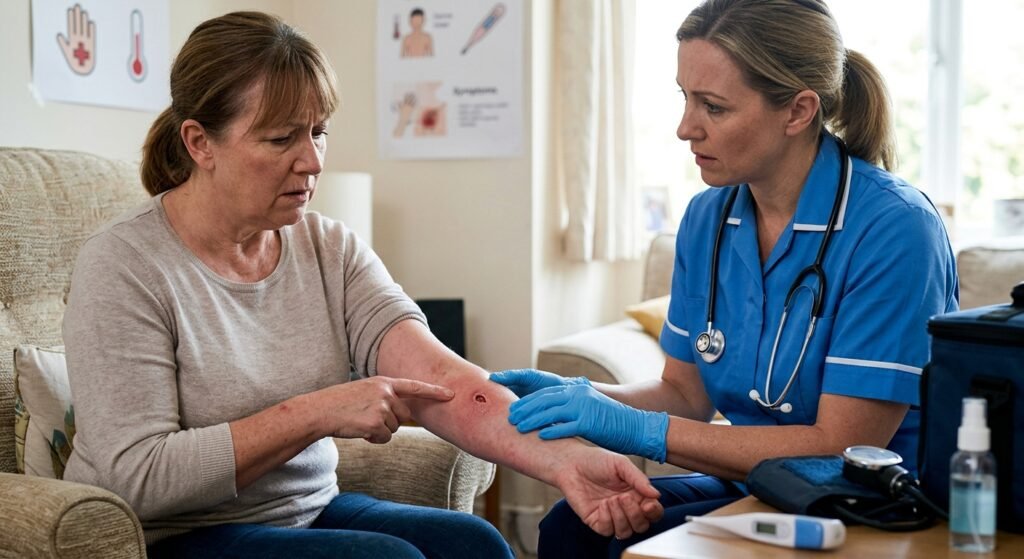
While self-monitoring using the ABCDE rule is valuable, nothing substitutes for a professional dermatologist exam. A trained dermatologist has years of experience recognizing subtle signs of skin cancer that the untrained eye might miss. They can perform a skin biopsy if needed, which is the definitive diagnostic test to confirm whether a lesion is cancerous or benign. Many serious cases of skin cancer could have been prevented or treated more easily if caught during an early professional exam.
What to Expect During a Dermatologist Exam
During a typical dermatologist appointment, your doctor will examine your entire body, including areas you might not think to check yourself, like your scalp, between your toes, and under your nails. They’ll use specialized equipment to get a better look at concerning lesions, and they may take photographs for future comparison. If they spot anything suspicious, they’ll likely recommend a skin biopsy, which involves removing a small sample of tissue for laboratory analysis. This test is the gold standard for diagnosing skin cancer and takes only a few minutes.
Comparing Different Skin Cancer Types: A Quick Reference Guide
| Type of Cancer | Most Common Appearance | Typical Location | Growth Rate | Survival Rate (Early Detection) |
|---|---|---|---|---|
| Melanoma | Irregular mole with multiple colors, asymmetrical | Sun-exposed areas, can appear anywhere | Fast | 99% (Stage 1) |
| Basal Cell Carcinoma | Shiny bump, pink growth, scar-like lesion | Face, ears, neck | Slow | 99%+ |
| Squamous Cell Carcinoma | Scaly patch, wart-like growth, non-healing sore | Face, lips, hands | Slow to moderate | 95%+ |
| Actinic Keratosis | Scaly, rough patch, red or tan | Sun-exposed areas | Slow (precancerous) | N/A (precancerous) |
| Merkel Cell Carcinoma | Firm nodule, shiny button-like, red-purple | Face, neck, sun-exposed areas | Fast | 60-80% (varies by stage) |
Essential Do’s and Don’ts for Skin Cancer Prevention and Detection
| DO | DON’T |
|---|---|
| Do perform monthly self-examinations using a mirror | Don’t ignore mole changes or new growths |
| Do use SPF 30+ sunscreen daily, even on cloudy days | Don’t use tanning beds under any circumstances |
| Do wear protective clothing, hats, and sunglasses during peak UV hours | Don’t delay seeing a dermatologist if you notice suspicious lesions |
| Do get a professional skin exam at least annually | Don’t assume all moles look the same or should look the same |
| Do keep track of your moles by taking photos for comparison | Don’t squeeze, pick, or attempt to treat suspicious lesions at home |
| Do apply sunscreen generously and reapply every two hours | Don’t spend extended time in the sun between 10 AM and 4 PM |
| Do examine all areas of your body, including hard-to-see spots | Don’t rely solely on internet research to diagnose skin conditions |
| Do inform your dermatologist of any family history of skin cancer | Don’t postpone treatment once skin cancer is diagnosed |
Risk Factors You Should Take Seriously
Who Is Most Vulnerable to Skin Cancer?
Certain groups of people face elevated risk of developing skin cancer. Those with fair or pale skin, particularly individuals of Celtic or Scandinavian descent, have less melanin in their skin, which provides less natural protection from UV radiation. People with a family history of skin cancer, particularly melanoma, have significantly higher risk. Additionally, individuals over forty years old have had more cumulative sun exposure over their lifetime. People with a history of severe sunburns, especially during childhood, and those with a large number of moles on their body also face increased risk.
Immunosuppression and Other Medical Risk Factors
People with weakened immune systems, whether due to HIV/AIDS, organ transplants, or certain medications, face dramatically increased risk of developing skin cancer. Additionally, those with a personal history of skin cancer should be extra vigilant about monitoring for new lesions, as they’re at higher risk for developing additional cancers.
The Importance of Early Detection and Treatment
Why Catching Skin Cancer Early Changes Everything
The difference between skin cancer caught in its earliest stages and skin cancer that has progressed is literally the difference between life and death in many cases. When melanoma is caught before it spreads to lymph nodes, the five-year survival rate exceeds ninety-five percent. However, if melanoma spreads to other organs, this survival rate drops dramatically. This dramatic difference emphasizes why regular self-examination and professional dermatologist exams are so crucial.
Understanding Your Treatment Options
Treatment options for skin cancer vary depending on the type, stage, and location of the cancer. Early-stage basal cell carcinoma and squamous cell carcinoma are often treated with surgical removal, which has excellent cure rates. Melanoma treatment might involve surgery, immunotherapy, targeted therapy, or radiation depending on the stage. Actinic keratosis is typically treated with topical medications, cryotherapy, or laser therapy to prevent progression to squamous cell carcinoma. Your dermatologist will recommend the most appropriate treatment based on your specific situation.
Taking Action: Your Next Steps
Schedule That Overdue Dermatologist Appointment
If you’ve been putting off scheduling a dermatologist exam, today is the day to make that appointment. Don’t wait until a concerning lesion appears or until you notice significant mole changes. Regular professional evaluation is the best investment you can make in protecting your skin health. If you notice any suspicious skin lesions in the meantime, don’t hesitate to call your dermatologist’s office and ask for an urgent appointment.
Establish a Regular Self-Monitoring Routine
Set a monthly reminder to perform a thorough self-examination of your skin using the ABCDE rule. Take photographs of any moles or lesions you’re concerned about so you can track whether they change over time. Keep these photos in a folder where you can easily compare them month to month. This simple practice can help you catch changes early and provide valuable information to share with your dermatologist.
Make Meaningful Lifestyle Changes Today
Start protecting your skin immediately by applying sunscreen daily, wearing protective clothing, and avoiding peak sun hours whenever possible. Never use tanning beds, regardless of how safe they claim to be. If you have a family history of skin cancer, inform your healthcare providers and follow their recommendations for screening intervals. Share this information with family members and friends—skin cancer awareness can literally save lives.
Conclusion: Your Skin Health Is Worth the Effort
Recognizing what skin cancer looks like is one of the most powerful tools you have for protecting your health. By understanding the visual characteristics of melanoma, basal cell carcinoma, squamous cell carcinoma, and other dangerous lesions, you gain the ability to identify problems early when treatment is most effective. Combined with regular dermatologist exams and vigilant sun protection, this knowledge provides comprehensive protection against one of the most common and preventable cancers.
Your skin is your body’s largest organ, and it deserves your attention and care. Don’t ignore suspicious changes, don’t postpone professional evaluation, and don’t underestimate the importance of prevention. The few minutes you invest in monthly self-examination and the time you spend applying sunscreen are trivial compared to the peace of mind and health benefits you’ll gain. Start today—your future self will thank you.
2 Comments