

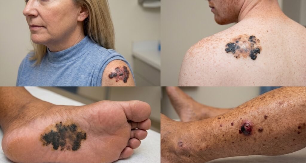
What Is Melanoma? If you’ve ever felt a flutter of fear after discovering an unusual mole on your body, you’re experiencing a concern shared by millions of people each year. Melanoma is a serious form of skin cancer and one of the most aggressive skin conditions that can develop on the skin. It begins in melanocytes, the specialized cells responsible for producing melanin, which gives your skin its natural color and helps protect it from harmful UV exposure. Every year, many people receive a melanoma diagnosis without fully understanding what this cancer means for their health and future.
Although melanoma accounts for a smaller percentage of overall skin cancer cases, it causes a significant proportion of skin cancer-related deaths because of its ability to grow and spread to other parts of the body. Unlike basal cell carcinoma and squamous cell carcinoma, which generally grow more slowly and are less likely to spread, melanoma can metastasize through the lymphatic system and bloodstream. Once melanoma spreads to distant organs, it becomes metastatic melanoma, which can be more challenging to treat.
Understanding What Melanoma Really Is: More Than Just a Skin Condition
The Basics: What Happens When Skin Cancer Becomes Melanoma
When you’re first told that you have melanoma, your immediate reaction is likely confusion mixed with fear. What exactly is this disease, and why is it so much more serious than other types of skin cancer? At its core, melanoma is a cancer that begins in the melanocytes—those special cells in your skin that produce melanin. Think of melanin as your skin’s natural sunscreen; it’s the pigment that darkens your skin when you get a tan and protects you from the sun’s harmful ultraviolet radiation. But when UV exposure damages the DNA within these melanocytes, something goes terribly wrong. The cells begin to mutate and multiply out of control, growing in a disorganized, abnormal way. This uncontrolled growth is what transforms a normal mole into a dangerous melanoma.
The reason melanoma is so feared in the medical community isn’t just because it grows aggressively. It’s because melanoma has a nasty habit of spreading. Unlike basal cell carcinoma that stays localized in your skin, melanoma cells can break away from the original tumor and travel throughout your body. When this happens, it becomes metastatic melanoma, and suddenly you’re dealing with a systemic disease rather than just a skin problem. This ability to spread is what makes early detection so critically important—catching melanoma when it’s still confined to the skin gives you the best possible chance at successful treatment.
How Melanoma Develops: The Cellular Story
Understanding how melanoma actually develops at the cellular level helps explain why this disease is so dangerous and why prevention is so crucial. Your skin contains thousands of melanocytes scattered throughout the deepest layer, called the basal layer. These cells exist in perfect harmony under normal circumstances, producing just enough melanin to protect you from the sun. But when UV radiation penetrates your skin, it damages the DNA inside your melanocytes. This damage is supposed to trigger your cells to repair themselves or self-destruct if the damage is too severe. However, sometimes the repair mechanisms fail, and the damaged cells survive with mutated DNA. Over time, especially with repeated sun exposure, these mutated cells accumulate, and eventually, they can transform into malignant melanoma.
The development of melanoma often involves specific genetic mutations that scientists have been studying intensely. The most common mutations include BRAF mutations, which are found in about fifty percent of melanoma cases, NRAS mutations, and CDKN2A mutations. There’s also the MC1R gene, which controls the production of red and yellow pigments in addition to dark melanin. People with certain variations of the MC1R gene, particularly those with red hair and fair skin, have a higher risk of developing melanoma because their skin produces less protective dark melanin. These genetic mutations can be inherited, meaning some people are born with a predisposition to melanoma, or they can be acquired during your lifetime as a result of sun damage.
The Different Types of Melanoma You Should Know About
Superficial Spreading Melanoma: The Most Common Form
When dermatologists talk about melanoma statistics, they mention that superficial spreading melanoma is responsible for about seventy percent of all cases. This form of melanoma develops horizontally across your skin in the early stages before it potentially grows deeper. The growth pattern is what gives this type its name—it spreads outward along the surface of your skin. Superficial spreading melanoma often appears as an irregular, multicolored lesion with an uneven border. The good news is that because it initially grows slowly across the skin surface, there’s a window of opportunity to catch it early before it penetrates deeper and becomes more dangerous.
Nodular Melanoma: The Aggressive and Deceptive Type
Nodular melanoma is a particularly aggressive form that presents a different challenge. Rather than spreading horizontally first, nodular melanoma grows downward into the deeper layers of skin from the very beginning. This vertical growth pattern means that nodular melanoma can penetrate deep into the dermis and beyond much faster than other types. Because of this aggressive behavior, nodular melanoma is often diagnosed at a more advanced stage. It typically appears as a raised bump or nodule that might be brown, black, blue, or even the color of normal skin. Some nodular melanomas don’t even have the darkly pigmented appearance people expect from melanoma, which can lead to delayed diagnosis. The fact that nodular melanoma can appear relatively innocuous while being extremely dangerous makes it one of the most concerning types.
Acral Lentiginous Melanoma: The Hidden Threat on Hands and Feet
Acral lentiginous melanoma is a type that affects the palms of your hands, the soles of your feet, or under your nails. The term “acral” refers to these distal extremities—the farthest points from your center body. This type of melanoma accounts for a smaller percentage of overall melanoma cases, particularly in lighter-skinned populations, but it’s notably more common in people with darker skin tones, Asian populations, and Indigenous populations. Acral lentiginous melanoma often goes undetected because people don’t routinely examine their palms and soles the way they might examine their arms or legs. When it appears on the nail, it might look like a dark streak or discoloration that people mistake for a bruise or stain under the nail. By the time acral lentiginous melanoma is discovered, it’s often at a more advanced stage, which underscores the importance of examining all areas of your body.
Lentigo Maligna: The Slow-Growing Threat on Sun-Damaged Skin
Lentigo maligna is a type of melanoma that develops on sun-damaged skin, typically in older adults who have had decades of cumulative sun exposure. It usually appears on the face, ears, or neck—areas that get constant sun exposure year-round. Lentigo maligna often looks like a large, irregular patch of brown, tan, or black pigmentation. What makes lentigo maligna different from other melanomas is that it can remain confined to the epidermis (the outermost layer of skin) for many years, sometimes even decades. However, the concerning aspect is that lentigo maligna has the potential to progress into lentigo maligna melanoma, a more aggressive form that invades deeper layers of skin. For this reason, dermatologists usually recommend treating lentigo maligna rather than simply monitoring it.
Amelanotic Melanoma: The Invisible Killer
Perhaps the most terrifying type of melanoma is amelanotic melanoma—a form that lacks the typical dark pigmentation people associate with melanoma. “Amelanotic” literally means “without melanin,” and these melanomas might appear pink, red, skin-colored, or even purple. Because amelanotic melanoma doesn’t look like what people expect melanoma to look like, it’s frequently missed or misdiagnosed. People might mistake an amelanotic melanoma for a harmless red bump, a scar, or a bloodspot. By the time it’s correctly identified, it has often progressed significantly. The danger of amelanotic melanoma emphasizes why professional evaluation by a dermatologist is so important—your naked eye and untrained assessment might miss something that a trained expert would immediately recognize as suspicious.
Recognizing Melanoma Using the ABCDE Rule
Asymmetry: When One Half Doesn’t Match the Other
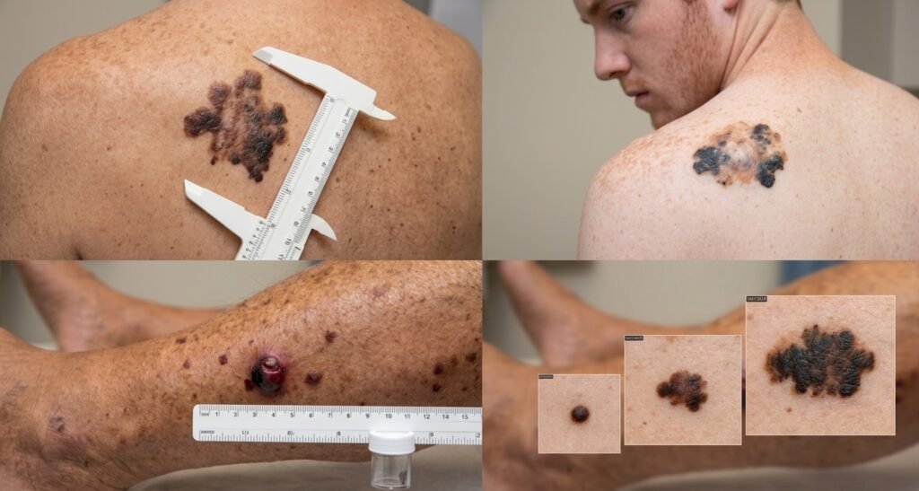
The ABCDE rule is a simple but powerful tool that can help you catch melanoma early. Let’s break it down, starting with “A” for asymmetry. To check for asymmetry, imagine drawing an invisible line down the middle of any mole or lesion on your skin. A benign mole is typically symmetrical—if you folded it in half, both sides would match. But melanoma is often asymmetrical, meaning one half looks completely different from the other. This asymmetry happens because melanoma grows in a chaotic, uncontrolled manner, whereas normal moles grow evenly.
Border Irregularity: The Ragged Edges That Signal Danger
The “B” in the ABCDE rule stands for border irregularity. Normal moles have clear, well-defined borders where the pigmented area ends and normal skin begins. Melanoma, by contrast, often has irregular, scalloped, or notched borders that look almost like a coastline with jagged peninsulas and inlets. The border might be blurry or fade gradually into the surrounding skin rather than having a clean line of demarcation. If you look closely and the edge seems uneven or wavy rather than smooth and consistent, this is a warning sign.
Color Variation: When Multiple Hues Appear in One Spot
The “C” stands for color, and specifically, color variation is what you’re looking for. A normal mole is usually one consistent color throughout—either brown, tan, or black. But melanoma often displays multiple colors within the same lesion. You might see black, brown, tan, red, pink, blue, or even white all mixed together in one area. The presence of three or more distinct colors, particularly if they seem irregular and scattered rather than evenly distributed, should raise red flags. This color variation reflects the chaotic nature of melanoma growth and the presence of cancer cells in different stages of development.
Diameter: The Six-Millimeter Guideline
The “D” in ABCDE refers to diameter. The rule of thumb is that any mole larger than six millimeters in diameter—roughly the size of a pencil eraser—warrants evaluation by a dermatologist. However, it’s important to understand that size alone isn’t an absolute rule. Some melanomas can be smaller than six millimeters, while some large moles might be completely benign. Size should be evaluated in combination with the other ABCDE criteria. If a mole is large and has other concerning features, that’s especially worrisome.
Evolution: The Most Important Warning Sign of All
The “E” stands for evolution, and many dermatologists consider this the most important letter in the entire rule. Evolution means change—any modification in a mole’s size, shape, color, or texture over time. If a mole that you’ve had for years suddenly starts growing larger, changing color, developing an uneven appearance, bleeding, or becoming itchy, this is a red flag. Melanoma doesn’t stay static; it changes over time. If you notice that a mole is different from how you remember it, or if it looks different from your other moles, this is definitely worth having professionally evaluated.
Understanding Melanoma Risk Factors and Genetic Predisposition
Sun Exposure and UV Radiation: The Primary Environmental Risk Factor
While melanoma can develop in people without significant sun exposure, UV exposure is still considered the primary environmental risk factor for developing this disease. Interestingly, it’s not just the total amount of sun exposure that matters—the pattern of exposure is also significant. People who experience intermittent, intense sun exposure with severe sunburns appear to have higher melanoma risk than people with chronic, continuous sun exposure. If you spent your childhood and teenage years getting sunburned at the beach during summer vacations, you’re at higher risk than someone who was gradually tanned year-round. This is why melanoma rates are actually highest in temperate climates where people vacation in the sun, not in equatorial climates where people are continuously exposed.
Genetic Predisposition: Born with Higher Risk
Some people are literally born with a higher risk of developing melanoma because of their genetics. Melanoma risk factors include a family history of melanoma, which increases your personal risk dramatically. If your parents or siblings have had melanoma, your risk is significantly elevated. Additionally, certain inherited genetic mutations make people more susceptible. The CDKN2A gene mutation, for instance, is associated with familial melanoma syndrome, and people who inherit this mutation have a much higher lifetime risk of developing melanoma. The BRAF mutation, while often acquired rather than inherited, can be present in some families with multiple melanoma cases.
Skin Type and Appearance Factors
People with fair or pale skin have higher melanoma risk than people with darker skin tones, primarily because they have less protective melanin. Red-haired individuals with their characteristic fair skin and the MC1R gene variations have particularly high risk. People with many moles on their body—particularly those with more than fifty moles or those with atypical moles (also called dysplastic nevi)—have higher melanoma risk. Having atypical moles means having moles that are larger, irregularly shaped, or multicolored compared to typical moles. These atypical moles can occasionally transform into melanoma, and they’re considered a warning sign that requires increased vigilance.
Age and Other Risk Factors
While melanoma can occur at any age, including in children, it’s more common in adults over thirty. However, melanoma is also one of the most common cancers in young adults and is increasingly being diagnosed in younger age groups. A personal history of severe sunburns, particularly during childhood, increases your risk. Immunosuppression—whether from HIV/AIDS, organ transplants, or immunosuppressive medications—significantly increases melanoma risk. Interestingly, people with certain occupations that require extensive outdoor work have higher rates of melanoma despite having darker tans.
Early Detection and Diagnosis: Your Best Defense Against Melanoma
How Melanoma is Diagnosed: From Suspicion to Confirmation
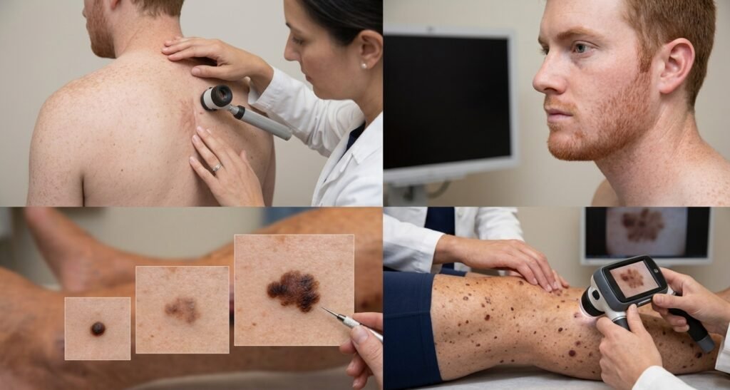
If you notice a concerning mole using the ABCDE rule, the next step is professional evaluation by a dermatologist. During your appointment, your dermatologist will examine the lesion closely, often using a tool called a dermoscope that magnifies the area and reveals patterns not visible to the naked eye. If the lesion looks suspicious, the dermatologist will recommend a biopsy. A biopsy involves removing all or part of the suspicious lesion and sending it to a laboratory for microscopic examination. A dermatopathologist—a specialist in skin pathology—will look at the cells under a microscope and determine whether melanoma is present and, if so, how advanced it is.
Staging: Understanding How Far the Melanoma Has Progressed
Once melanoma is diagnosed, it’s staged to determine how serious it is and what treatment options are appropriate. Early-stage melanomas that are thin and haven’t spread have excellent prognosis. But if the melanoma has grown thick or has already begun spreading to lymph nodes or distant organs, becoming metastatic melanoma, the situation is much more serious. Staging considers the thickness of the melanoma, whether it has spread to lymph nodes, and whether it has metastasized to distant organs. This staging information guides all treatment decisions.
Genetic Testing and Mutation Analysis
Modern melanoma diagnosis often includes testing for specific genetic mutations. If your melanoma has a BRAF mutation, this information is important because it affects which treatments might work best for you. Some targeted therapies specifically target BRAF-mutated melanomas. Similarly, testing for NRAS and other mutations can guide treatment decisions. Genetic counseling might be recommended if you have a strong family history of melanoma, particularly if you have multiple relatives affected at young ages.
Treatment Approaches: From Surgery to Immunotherapy
Surgical Treatment: The Foundation of Melanoma Care
Surgery is the primary treatment for most melanomas, particularly those caught early. The surgeon will remove the melanoma along with a margin of surrounding healthy tissue. How much surrounding tissue is removed depends on the thickness and characteristics of the melanoma. Early-stage melanomas might require only a small surgical margin, while thicker melanomas require wider removal. If your melanoma has spread to lymph nodes, sentinel lymph node biopsy might be performed during surgery to determine if the cancer has spread beyond the skin.
Immunotherapy: Harnessing Your Immune System to Fight Cancer
One of the most exciting developments in melanoma treatment is immunotherapy—medications that help your own immune system recognize and attack cancer cells. Checkpoint inhibitor drugs like pembrolizumab and nivolumab work by removing the “brakes” that melanoma cells use to hide from your immune system. These immunotherapy drugs have transformed the prognosis for people with metastatic melanoma, extending survival significantly for some patients. Immunotherapy can be used alone or in combination with other treatments.
Targeted Therapy for BRAF-Mutated Melanoma
If your melanoma has a BRAF mutation, you might be a candidate for targeted therapy drugs that specifically attack cells with this mutation. These drugs work by blocking the faulty BRAF protein, effectively shutting down the cancer cells’ growth signals. Targeted therapy can provide rapid response in BRAF-mutated melanomas, though resistance can develop over time.
Radiation and Systemic Treatments
For metastatic melanoma that has spread to the brain or other distant organs, radiation therapy might be used. Chemotherapy, while less commonly used now due to the success of newer immunotherapies, might still be considered in certain situations. Clinical trials might offer access to newer treatments not yet available through standard care.
Living with Melanoma: Prognosis, Surveillance, and Life After Diagnosis
Understanding Melanoma Prognosis: What Does Your Diagnosis Mean?
The word “prognosis” refers to the expected course and outcome of your disease. For melanoma, prognosis depends heavily on how early the cancer was caught. If your melanoma was caught at Stage 1 or 2, when it’s still confined to the skin, your prognosis is excellent—the five-year survival rate exceeds ninety percent. However, if your melanoma has spread to lymph nodes (Stage 3) or distant organs (Stage 4, metastatic melanoma), the outlook is more serious. But even metastatic melanoma can now be managed much more effectively than it could be just a decade ago, thanks to advances in immunotherapy. Many people with metastatic melanoma are now living longer and enjoying better quality of life than previously possible.
The Importance of Surveillance After Treatment
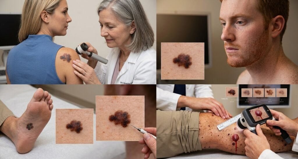
After melanoma treatment, careful surveillance becomes essential. This means regular dermatologic exams to look for recurrence of the original melanoma or development of new melanomas. People who have had melanoma have a higher risk of developing a second melanoma. Your dermatologist might recommend exams every three months initially, with spacing extended over time if no recurrence is detected. You’ll also be taught to perform monthly self-examinations using the ABCDE rule. Taking photographs of your moles to track changes over time can be incredibly helpful for both you and your dermatologist.
Psychological Impact and Support
Receiving a melanoma diagnosis can be psychologically devastating, even for early-stage cases. Many people experience anxiety, depression, and existential concerns about mortality. Support groups, both in-person and online, connect you with others who have experienced melanoma and understand what you’re going through. Talking with a therapist or counselor who specializes in cancer-related concerns can help you process your diagnosis and develop coping strategies. Don’t underestimate the emotional impact of melanoma—addressing your mental health is just as important as addressing the physical disease.
Comparing Melanoma Types: Essential Reference Guide
| Melanoma Type | Frequency | Growth Pattern | Typical Appearance | Common Location | Prognosis |
|---|---|---|---|---|---|
| Superficial Spreading | 70% of cases | Horizontal first, then deep | Irregular, multicolored, asymmetrical | Any sun-exposed area | Best (early detection possible) |
| Nodular | 15-30% of cases | Vertical from start | Raised bump or nodule | Any area of body | More serious (aggressive growth) |
| Acral Lentiginous | 5-10% of cases | Horizontal, then deep | Dark streak under nail or palm/sole patch | Palms, soles, under nails | More serious (often late diagnosis) |
| Lentigo Maligna | 4-15% of cases | Horizontal/slow | Large irregular tan/brown patch | Face, ears, neck | Variable (depends on progression) |
| Amelanotic | 5-10% of cases | Rapid | Pink, red, or skin-colored | Any area | More serious (often missed initially) |
Melanoma Prevention and Protection: What You Can Actually Control
| Prevention Strategy | Why It Matters | How to Implement |
|---|---|---|
| Daily Sunscreen Use | Prevents UV damage to melanocytes and reduces melanoma risk | Apply SPF 30+ every morning; reapply every 2 hours if outdoors |
| Avoid Peak Sun Hours | UV radiation is strongest between 10 AM and 4 PM | Plan outdoor activities before or after these hours |
| Protective Clothing | Physical barrier blocks UV rays better than sunscreen alone | Wear long sleeves, pants, hats, and UV-blocking sunglasses |
| Regular Self-Exams | Early detection dramatically improves outcomes | Perform monthly full-body exams using ABCDE rule |
| Professional Exams | Dermatologists catch melanomas you might miss | Annual exam recommended; more frequent if high risk |
| Avoid Tanning Beds | Artificial UV radiation increases melanoma risk | Never use tanning beds regardless of marketing claims |
| Family Communication | Genetic predisposition means relatives are at risk too | Share melanoma history with family; encourage their vigilance |
| Education and Awareness | Knowledge empowers you to recognize danger signs | Stay informed about melanoma signs and risk factors |
Essential Lifestyle Modifications After Melanoma Diagnosis
Sun Protection: A New Way of Life
If you’ve been diagnosed with melanoma, sun protection becomes non-negotiable. You need to approach sun safety with the same diligence you’d apply to taking essential medications. This means applying broad-spectrum SPF 30 or higher sunscreen every single day, even on cloudy days and even if you’re staying indoors most of the day. Reapply every two hours if you’re outside, and more frequently if you’re swimming. Wear protective clothing, including long sleeves and pants when possible, wide-brimmed hats, and UV-blocking sunglasses. Seek shade whenever you can, and be especially vigilant during summer months when UV radiation is most intense.
Body Monitoring: Becoming Your Own Vigilant Observer
You know your body better than anyone else does. You notice small changes that might not be obvious to someone examining you for fifteen minutes in a doctor’s office. This is why monthly self-examinations are so important. Set a calendar reminder for the same day each month. Using a mirror and a handheld mirror for hard-to-see areas, examine your entire body systematically. Look at your face, scalp (part your hair to check), ears, neck, arms, torso, legs, and feet. Don’t forget easily overlooked areas like between your toes, your armpits, and your buttocks. Take photographs of any moles that concern you, and compare them month to month. Any changes—in size, color, shape, or how the mole feels—warrant professional evaluation.
Regular Dermatologic Follow-Up: Your Safety Net
After melanoma treatment, you’ll need regular dermatologic follow-up, typically every three months initially. These appointments aren’t just about looking for melanoma recurrence on your skin—they’re also about monitoring for systemic signs of metastatic melanoma. Your dermatologist might recommend blood tests or imaging studies depending on your specific situation. Don’t skip these appointments, even when you’re feeling perfectly fine. Many recurrences are caught through scheduled surveillance rather than because you noticed symptoms.
The Reality of Living with Melanoma Awareness
Moving Beyond Fear to Empowerment
Learning that you have melanoma, or learning that you’re at high risk for melanoma, can feel like living under a dark cloud. The fear is real, and it’s understandable. But knowledge is power. By understanding what melanoma is, recognizing the warning signs, knowing your risk factors, and understanding your treatment options, you transform yourself from a passive victim of disease into an active participant in your health care. This shift in perspective doesn’t make the fear disappear, but it channels that fear into productive action.
Hope in Modern Medicine
Just a few decades ago, a melanoma diagnosis, particularly a metastatic melanoma diagnosis, was often a death sentence. Today, thanks to immunotherapy, targeted therapy, and other advances in medical science, many people with melanoma are living longer and enjoying better quality of life than ever before. New treatments are being developed constantly, and clinical trials offer access to cutting-edge therapies. While melanoma remains a serious disease that demands respect and vigilance, the hopelessness that once accompanied a melanoma diagnosis has been replaced by genuine optimism.
Conclusion: Taking Control of Your Melanoma Risk
Melanoma is a serious disease that demands your attention and respect, but it’s not an inevitable part of life. The combination of sun protection, regular self-examination using the ABCDE rule, professional dermatologic evaluation, and awareness of your personal risk factors gives you powerful tools to either prevent melanoma altogether or catch it at the earliest, most treatable stage. Whether you’re trying to protect yourself from ever developing melanoma, or whether you’ve already received a diagnosis and are navigating treatment and surveillance, remember that you’re not alone. Thousands of dermatologists, oncologists, nurses, and support groups are committed to helping people prevent, detect, and treat melanoma effectively.
Start today by protecting your skin from UV exposure, scheduling an appointment with a dermatologist if you haven’t had one recently, and educating yourself about melanoma risk factors and warning signs. Your skin health is worth the investment of time and attention. By taking control now, you’re investing in a healthier, longer life.
1 Comment